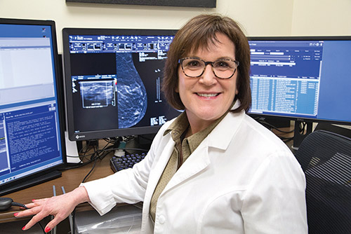
Ask the Doctor with Dr. Mindy Goldfischer
October is National Breast Cancer Awareness Month—a great time for women of all ages to learn more about breast health and double their efforts in surveilling for irregularities in their breast tissue.
Who is at high risk?
Most women who develop breast cancer have no known risk factors. Only 5-10% of breast cancers occur in women with a genetic mutation, usually BRCA1 or BRCA2.
A few high-risk factors to keep in mind:
History of breast cancer.
Having a genetic mutation or a first-degree relative with a genetic mutation. Women of Ashkenazi Jewish descent are at increased risk for BRCA mutation, as well as other less common mutations, such as CHEK2.
Family history (maternal or paternal) of breast cancer, especially in first-degree relatives. Age at diagnosis is important, with premenopausal occurrence increasing risk.
Chest radiation before the age of 30.
Having one or more breast biopsies for ADH (atypical ductal hyperplasia) or LCIS (lobular carcinoma in situ).
At what age should women begin breast cancer screening?
The American College of Radiology (ACR) recommends that women with an average risk of developing breast cancer begin annual screening mammography at age 40. Because younger women tend to have dense breast tissue, which can obscure masses, annual mammograms are important; sometimes supplemental breast ultrasound is indicated. Women at high risk should begin annual screening 10 years earlier, but not before the age of 25. After the age of 75, women should consult their physicians to determine whether they need additional screening.
What are the advantages of 3D mammography?
At Englewood Health, all mammograms are 3D. With the traditional 2D mammogram, the X-ray tube is stationary and the breast tissue overlaps in the image. With a 3D Mammography™ exam, the X-ray tube moves in an arc around the breast. Images are obtained from multiple angles and synthesized by a computer, which creates thin slices that can be viewed individually. A special computer algorithm achieves 3D mammograms with the same radiation dose as 2D.
Mindy Goldfischer, MD, is chief of breast imaging at The Leslie Simon Breast Care and Cytodiagnosis Center at Englewood Health.